MthK Ca2+ Activated Crystal Structure

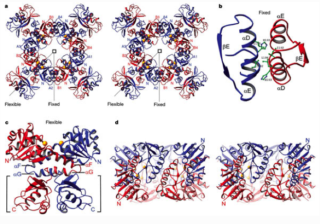
a, Stereo diagram of the gating ring viewed down the four-fold axis (square). Eight RCK domains are divided into two groups, A1–A4 (blue) and B1–B4 (red), with N termini labelled (N). Viewed from the membrane, blue domains would be attached to the pore via a continuous polypeptide chain and red domains assembled from solution. Fixed and flexible dimer interfaces hold the ring together. b, Helices D and E form the fixed interface. Several amino acids conserved as hydrophobic or aromatic (Ile 192, His 193 and Leu 196) are shown. c, C-terminal subdomains (brackets) and helices F and G form the flexible interface. A cleft between two RCK domains creates a ligand-binding site with Ca2+ bound at the base (yellow spheres). d, Stereo diagram of the gating ring viewed from the side after removing the subdomains.

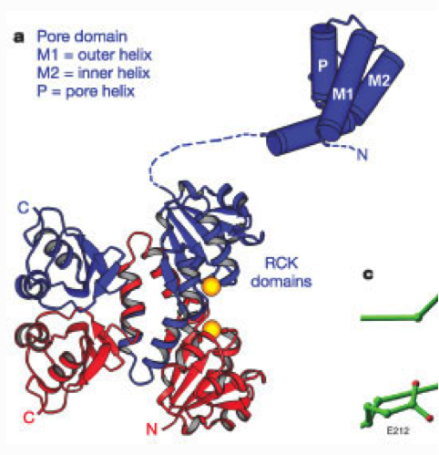
a, A single four-fold repeated unit of the MthK channel contains a pore domain (blue cylinders) and two RCK domains (blue and red ribbons). Disordered segments (dashed) and Ca2+ ions (yellow spheres) are shown
Jiang, Y., Lee, A., Chen, J., Cadene, M., Chait, B.T., MacKinnon, R. (2002). Crystal structure and mechanism of a calcium-gated potassium channel. Nature, 417, 515-522. doi: 10.1038/417515a
