Kv1.2 Crystal Structure

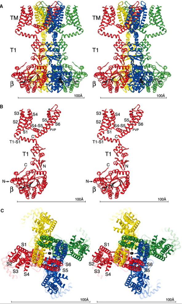
Views of the Kv1.2–β2 subunit complex. (A) Stereoview of a ribbon representation from the side, with the extracellular solution above and the intracellular solution below. Four subunits of the channel (including the T1 domain, voltage sensor, and pore) are colored uniquely. Each subunit of the β subunit tetramer is colored according to the channel subunit it contacts. The NADP+ cofactor bound to each β subunit is drawn as black sticks. TM indicates the integral membrane component of the complex. (B) Stereoview of a single subunit of the channel and β subunit viewed from the side. Labels correspond to transmembrane helices (S1 to S6); the Pro-Val-Pro sequence in S6 (PVP); and the N (N) and C (C) termini of the Kv1.2 and β subunits. The position of the N terminus of the β subunit, which is located on the side furthest away from the viewer, is indicated by an arrow. (C) Stereoview of a ribbon representation viewed from the extracellular side of the pore. Four subunits are colored uniquely.
Long, S., Campbell, E.B., MacKinnon, R. (2005). Crystal Structure of a Mammalian Voltage-Dependent Shaker Family K+ Channel. Science, 309(5736), 897-903. doi: 10.1126/science.1116269
