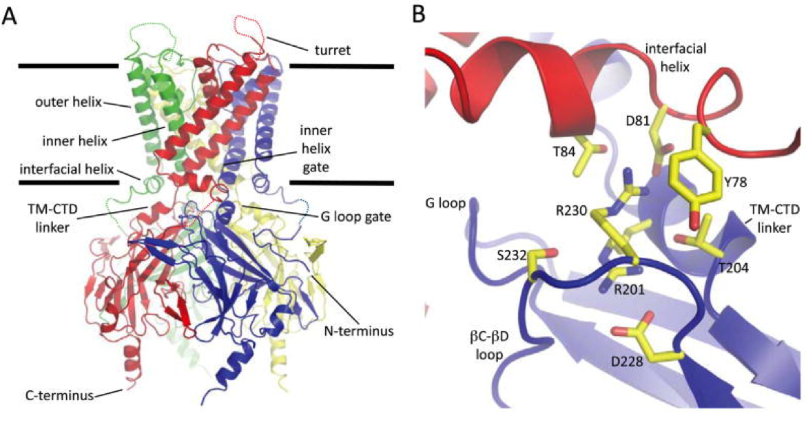
GIRK2 Crystal Structure

(A) Cartoon diagram of the GIRK2 structure. Each subunit of the tetramer is a different color. Unmodeled segments of the turret and N-terminal linker are drawn with dashed lines. The approximate boundary of the phospholipid bilayer is indicated by the thick black lines.
(B) A cartoon diagram of key residues that mediate the contacts at the interface between the cytoplasmic and transmembrane domains. The same coloring scheme as in panel A is used.
Whorton, M.R., MacKinnon, R. (2011). Crystal structure of the mammalian GIRK2 K+ channel and gating regulation by G-proteins, PIP2 and sodium. Cell, 147(1), 199-208. doi: 10.1016/j.cell.2011.07.046
