Active amplification through critical dynamics in an excised segment of the mammalian cochlea
Hearing benefits from the ear’s ability to enhance its responsiveness by means of an energy-expending active process. This process is defined by four properties that, although seemingly unrelated, consistently occur together in vertebrates: amplification, sharp frequency tuning, compressive nonlinearity, and spontaneous otoacoustic emission. In nonmammal tetrapods, this active process is evident in individual hair cells. The motile hair bundles of the bullfrog, for example, exhibit all four attributes by operating near a Hopf bifurcation—a critical regime in which these properties naturally coalesce. In mammals, however, the delicate nature of the cochlea has restricted the evidence for an active process to studies in vivo, where it is generally attributed to the collective effort of the outer hair cells that energize the traveling wave along the cochlear spiral. As a result, the cellular mechanisms that underlie the properties of mammalian hearing remain contested, with uncertainty about whether criticality plays a role in the cochlea's active process and even whether an active process exists. We have demonstrated the presence of an active process in an excised segment of the gerbil's cochlea in the absence of a traveling wave. We show that a cochlear segment displays amplification, frequency tuning, and compressive nonlinearity and can generate distortion products. By freeing the active process from the constraints of traveling waves, we show that mammalian hair cells, like those of other tetrapods, achieve active amplification through a dynamical system operating at criticality. Derived from a novel experimental system that allows optical and electrical examination as well as direct pharmacological manipulation, the results constitute a step toward unraveling the complexity of cochlear function. Our findings underscore the existence of a fundamental dynamical principle that underpins auditory processes across a range of vertebrate and invertebrate species.
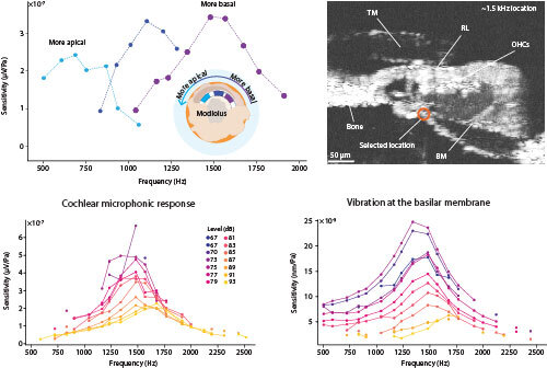
Upper left: For cochlear segments isolated from different locations along the cochlear spiral, which is shown schematically in a view from the apex, the sensitivity of the cochlear microphonic potential peaks at distinct frequencies. Upper right: An optical coherence tomogram (OCT) depicts a living organ of Corti near the 1.5 kHz tonotopic location. Lower left: At the 1.5 kHz position, the sensitivity of the cochlear microphonic response to frequency complexes at different intensities displays striking nonlinearity: the cells are far most sensitive to weak than to strong stimuli. Lower right: Similar behavior is observed for the sensitivity of basilar-membrane vibration at the position marked in red in the OCT.