Graphical Simulations
 |
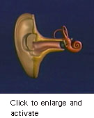
Cochlear traveling wave Sound, which consists of pressure changes in the air, is captured by the external ear, enters the ear canal, and vibrates the eardrum (tympanum) and the tiny associated bones (ossicles) of the middle ear: the hammer (malleus), anvil (incus), and stirrup (stapes). The last acts as a piston that produces pressure changes in the liquid-filled, snail-shaped inner ear (cochlea). This input sets the elastic basilar membrane (unrolled in the simulation for ease of visualization) into oscillation. Because the top end (apex) of the basilar membrane is broad and soft, it responds best to low-frequency stimuli; the bottom end (base) is narrow, taut, and thus most sensitive to high pitches. When a complex sound such as J. S. Bach's Toccata and Fugue in D Minor is heard, each constituent musical tone excites the basilar membrane in a specific place. The actual movements are much faster, at the frequencies of the tones, and far smaller, of atomic dimensions. Movements of the basilar membrane are detected by some 16,000 hair cells situated along its length; their electrical responses trigger firing in nerve fibers that communicate information to the brain. © 1997 Howard Hughes Medical Institute. This animation may be used, copied, and distributed, without modification and with the foregoing copyright notice included, for non-commercial scientific or educational purposes only; any other use requires the written permission of Howard Hughes Medical Institute (contact bricken@hhmi.org). Download QuickTime Movie |
