KCNQ1/CaM Structure
Structure of KCNQ1/CaM complex
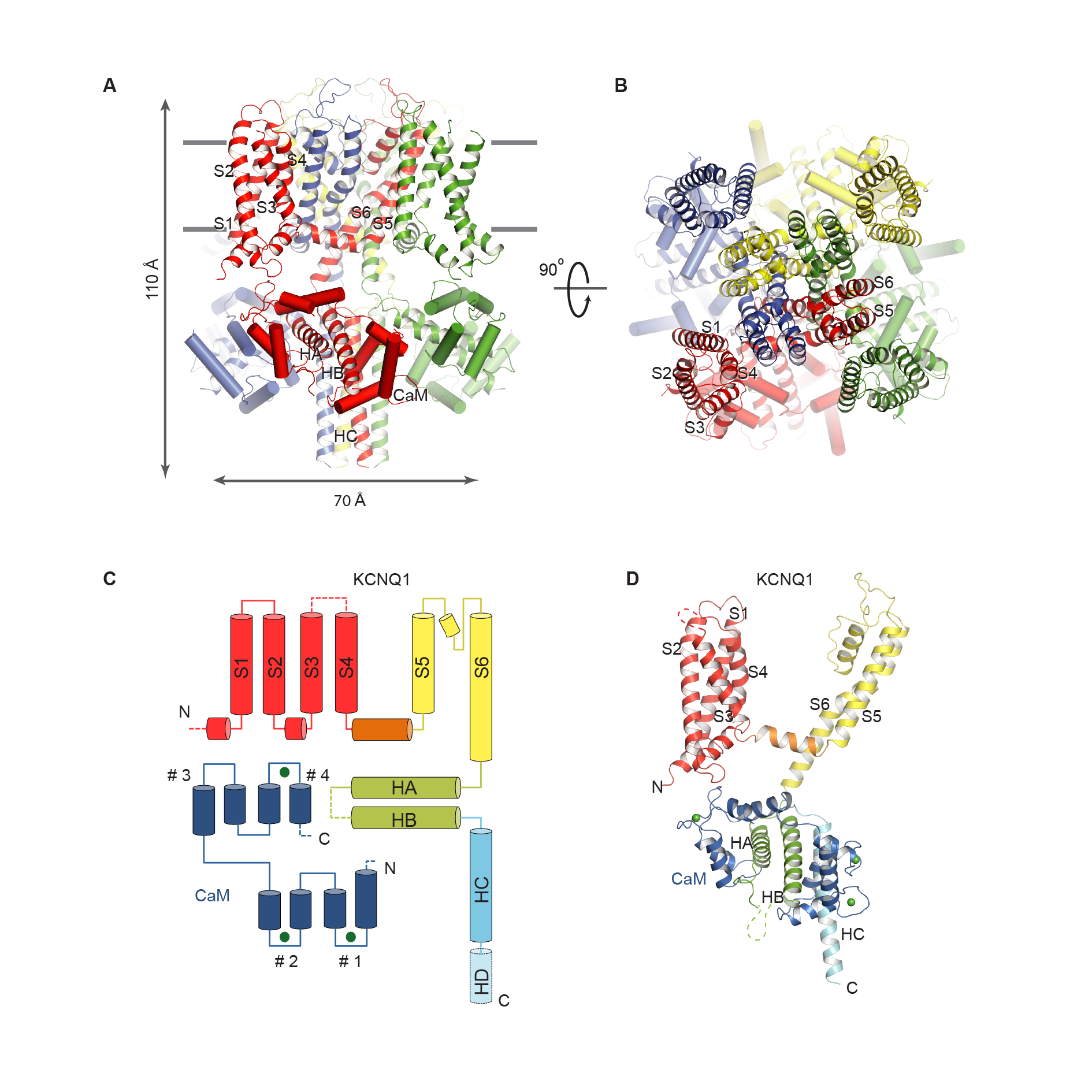
(A-B) Side view and top view of the KCNQ1/CaM complex. Each protomer is shown in a different color, and CaM is represented as cylinders. The S1-S6 and HA-HC are labeled.
(C) Domain organization of one protomer. The HD, which is masked out in 3D reconstruction, is indicated by cylinder with dashed outlines. The EF hands of CaM are labeled as #1 - #4 from the N-terminal to C-terminal end. The first two EF-hand regions form the N-lobe, and the other two form the C-lobe. Green spheres represent calcium ions.
(D) Model of one subunit with domains colored as in (C).
Sun, Ji, MacKinnon, R. (2017). Cryo-EM structure of a KCNQ1/CaM complex reveals insights into congenital long QT syndrome. Cell, 169(6), 1042-1050. PMID: 28575668.